Abstract Type : Oral presentation
Abstract Submission No.: A-0706
Abstract Topic : Transplantation
Lipid-Associated Macrophage Polarization and APOESDC2 Signaling Underlie Chronic Xenograft Fibrosis
Hyejin Jung1, Gaeun Byeon2, Minsun Jung3, Beom Seok Kim1, Joon-Yong An2, Jaeseok Yang1
1Department of Internal Medicine, Yonsei University College of Medicine, Korea, Republic of
2Department of Department of Integrated Biomedical and Life Science, Korea University, Korea, Republic of
3Department of Pathology, Yonsei University College of Medicine, Korea, Republic of
Objectives : Chronic injury of kidney xenografts, characterized by interstitial fibrosis and tubular atrophy (IF/TA), remains a primary barrier to long-term survival in pig-to-nonhuman primate (NHP) xenotransplantation. The molecular mechanisms driving this transition toward chronic rejection are poorly understood. In this study, we aimed to identify the cellular and molecular mechanisms underlying chronic xenograft fibrosis.
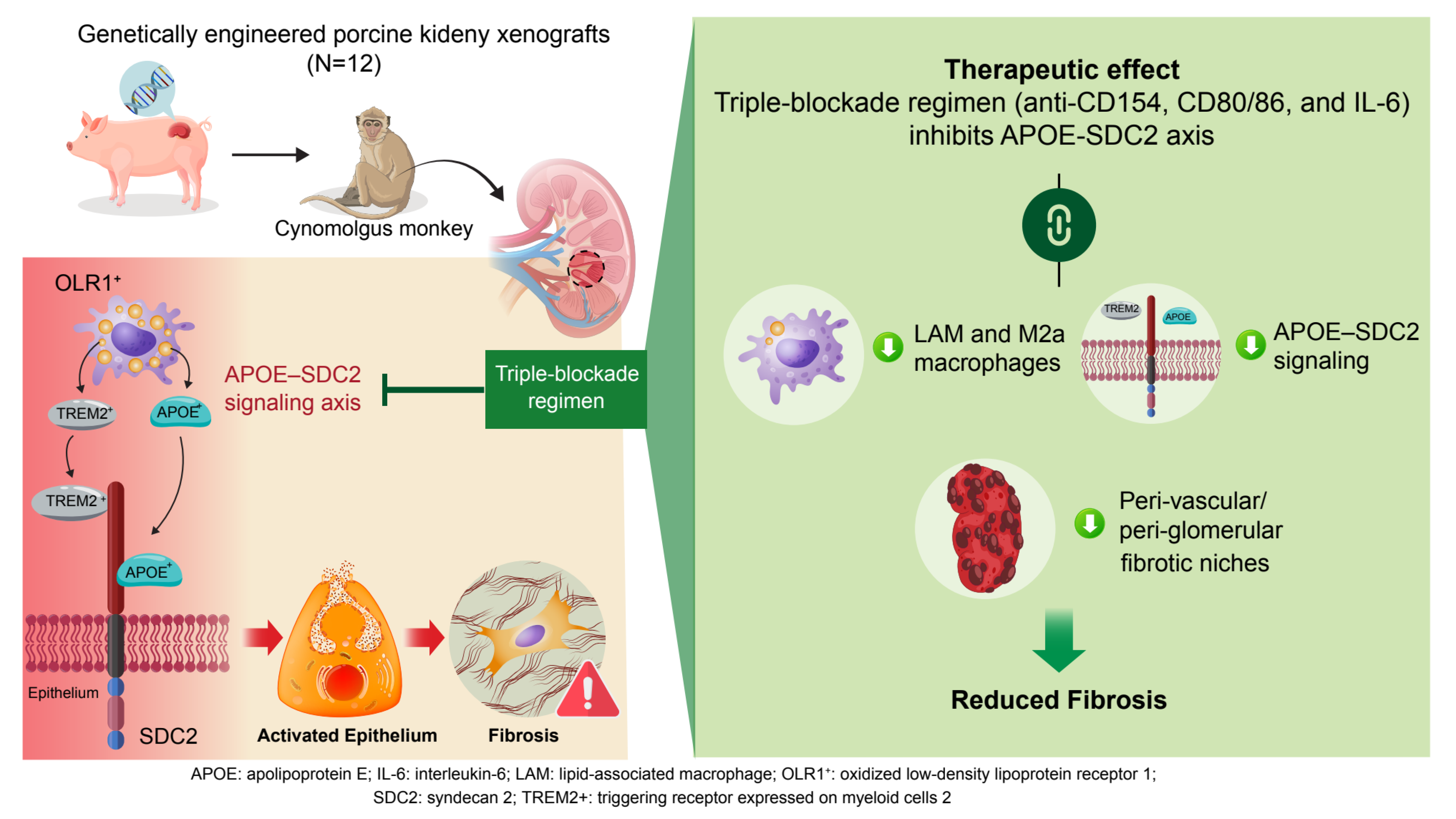
Methods : Kidney xenografts from genetically engineered porcine donors with 5–8 genetic modifications (knockouts for 3 or 4 major xenoantigens [GGTA1, CMAH, B4GalNT2 ± iGb3s] and knockins for 2 or 4 human complement- or coagulation-regulatory genes [CD39, CD55, CD46, TBM]) were transplanted into cynomolgus monkeys (n = 12) under five immunosuppressive regimens. They were assessed using a multi-modal transcriptomic approach, integrating single-nucleus RNA sequencing, spatial transcriptomics, and bulk RNA-sequencing, across multiple time points. Histopathology and Banff scoring were used to assess graft injury and fibrosis.
Results : We identified that increasing Banff fibrosis scores (ci [interstitial fibrosis] and ct [tubular atrophy]) correlate with macrophage polarization toward pro-fibrotic lipid-associated macrophage (LAM) and M2a phenotypes, driving tubulointerstitial fibrosis progression. Spatial transcriptomics further delineated pro-fibrotic immune–fibroblast and immune-adaptive epithelium niches enriched near glomeruli and arterioles. Notably, the APOE–SDC2 signaling axis emerged as a conserved mediator of macrophage-driven epithelial-mesenchymal transition (EMT). Furthermore, a triple-blockade regimen targeting CD154, CD80/86, and IL-6 signaling significantly mitigated fibrosis by dismantling these pro-fibrotic niches and suppressing the APOE–SDC2 axis.
Conclusions : Our study delineates the transcriptional and spatial landscape of chronic kidney xenograft injury and identifies distinct macrophage phenotypes and pro-fibrotic niches as central drivers of fibrosis. The therapeutic effect of targeted immunosuppression in mitigating the fibrotic processes highlights the potential of modulating immune–stromal interactions to improve long-term xenograft outcomes.
Figure.png